
The plaque your stress test never saw
A normal stress test rules out flow limiting narrowing, not the softer plaque where most heart attacks actually begin.
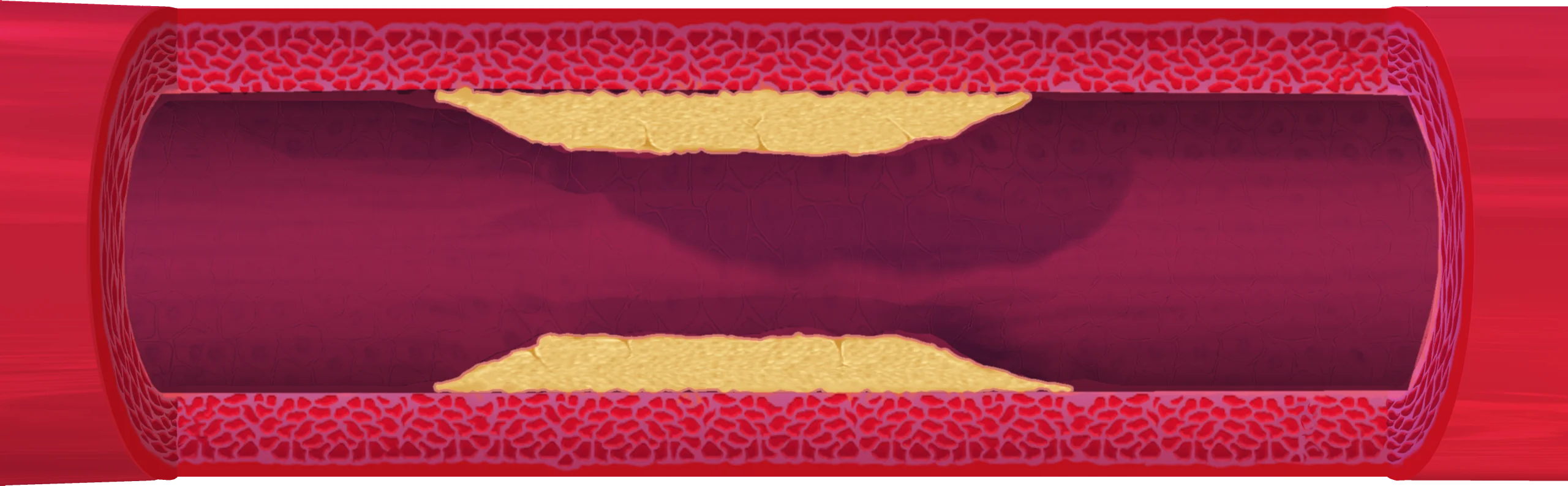 Soft plaque in the wall
Stress test: flow normal
A normal stress test is one of the most reassuring sentences in medicine, and one of the most misunderstood. It answers a narrower question than most people think, and the gap between that question and the one you actually care about is where a great many heart attacks begin.
What the treadmill is actually asking
A stress test looks for flow limitation. It asks whether a narrowing is tight enough that, when your heart works harder, some region of muscle cannot get the blood it needs. It answers that question well. It is a different question from whether there is atherosclerosis in the artery wall at all. Plaque can accumulate for years while the vessel remodels outward and keeps its channel open, so flow at peak exercise stays normal.
A normal stress test tells you nothing was flow limiting that day. It does not tell you your arteries are clean.
Where heart attacks actually start
Most heart attacks do not come from a narrowing that slowly closes off. They come from a plaque whose fibrous cap tears or erodes, with a clot forming on that spot. The plaque underneath is often modest, the kind that would not produce an abnormal stress test.

The clearest look at this came from the PROSPECT study, published in 2011. Nearly 700 patients had all three coronary arteries imaged with intravascular ultrasound after an acute coronary syndrome, then were followed for about three years. About half of the later cardiac events traced not to the artery that had been treated but to other lesions, and on the original angiogram those lesions had looked only mildly narrowed, averaging about a third of the vessel diameter.1 Most of those events were hospitalizations for worsening angina rather than heart attacks, which is the fairest criticism of the study. What marked the lesions was still not tightness. It was a large burden of plaque, a small lumen, and a thin cap.
33%
of diameter
A Veterans Affairs analysis in JAMA made the clinical version of the point. Among more than 37,000 patients sent for elective angiography, those with plaque that never reached the threshold we call obstructive still carried a higher one year risk of heart attack than those with no disease found.2 The excess was clearest once more than one vessel was involved. The absolute numbers at one year were small. The direction was not ambiguous.
This is why coronary CT angiography has moved toward the front of the workup. In the SCOT-HEART trial, adding CT to standard care for stable chest pain lowered the five year rate of coronary death or nonfatal heart attack from 3.9 percent to 2.3 percent, without more stents or bypass surgery.3 The most likely explanation is that more people were started on preventive treatment. The 2021 American Heart Association and American College of Cardiology chest pain guideline now gives CT angiography its strongest class of recommendation in several chest pain settings.4
None of this makes the stress test a bad test. It makes it a specific one. Everything above comes from people who had symptoms or had already had an event, so none of it is an argument for scanning everyone. It is an argument for knowing what your test did and did not rule out. If yours was normal and you still carry real risk, the question worth raising is not whether you passed, but what your overall risk looks like and whether any further imaging would change what you are treated with. That is a conversation for your own cardiologist, who knows your numbers and your history.
References
- Stone GW, et al. A prospective natural-history study of coronary atherosclerosis. New England Journal of Medicine, 2011;364(3):226-235 Read the source
- Maddox TM, et al. Nonobstructive coronary artery disease and risk of myocardial infarction. JAMA, 2014;312(17):1754-1763 Read the source
- SCOT-HEART Investigators. Coronary CT Angiography and 5-Year Risk of Myocardial Infarction. New England Journal of Medicine, 2018;379(10):924-933 Read the source
- Gulati M, et al. 2021 AHA/ACC/ASE/CHEST/SAEM/SCCT/SCMR Guideline for the Evaluation and Diagnosis of Chest Pain. Circulation, 2021;144(22):e368-e454 Read the source
Education, not medical advice.
Take any decision about your own heart with the clinician who knows your history.
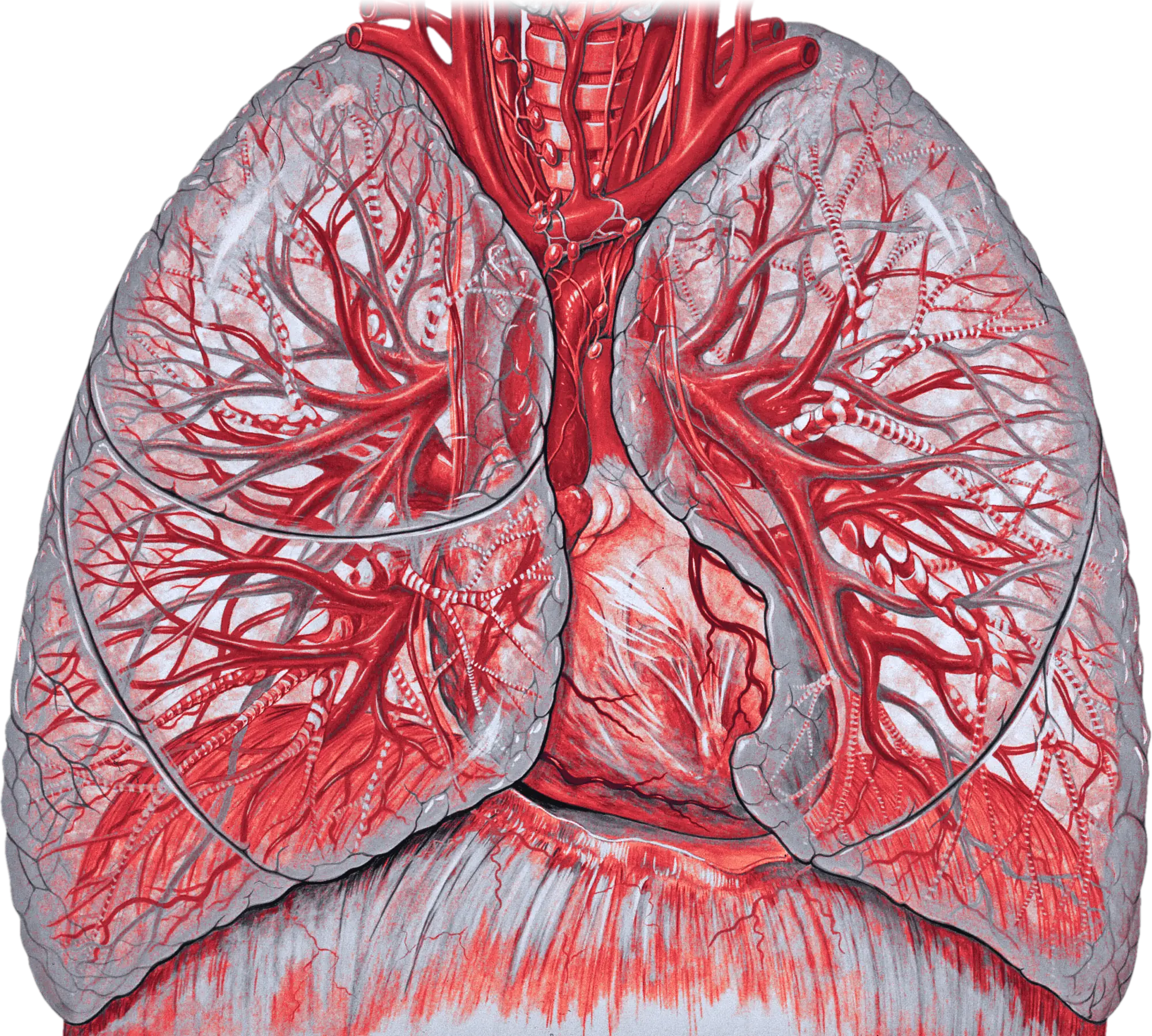
Illustration: Patrick J. Lynch and C. Carl Jaffe, MD, Yale University, CC BY 2.5 (adapted).